Home » Without Label » Knee Muscle Anatomy Mri - Supplemental Materials for Normal MR Imaging Anatomy of ... : When a muscle has different orientations of the tendons it means that there are different patterns of edema possible depending on the tendon injured.
Knee Muscle Anatomy Mri - Supplemental Materials for Normal MR Imaging Anatomy of ... : When a muscle has different orientations of the tendons it means that there are different patterns of edema possible depending on the tendon injured.
Knee Muscle Anatomy Mri - Supplemental Materials for Normal MR Imaging Anatomy of ... : When a muscle has different orientations of the tendons it means that there are different patterns of edema possible depending on the tendon injured.. The common peroneal nerve typically courses downward within abundant fat posterior to the short head of the biceps femoris muscle and superficial to the lateral head of the gastrocnemius muscle, but. In one investigation, depicted only on the proton density weighted images. The muscles of the knee include the quadriceps, hamstrings, and the muscles of the calf. Related posts of muscle anatomy knee mri muscle anatomy get body smart. Magnetic resonance imaging is particularly well suited for the medical evaluation of the musculoskeletal (msk) system including the knee, shoulder, ankle, wrist and elbow.
Superiorly, it extends to the level of the crossing of the biceps femoris tendon, and remains superficial to fcl in this location.10 The knee joint is a modified hinge joint between the femur tibia and patella. Plantaris acts weakly to plantar flex the foot and flex the knee. When interpreting the proton density images it. Anatomy basic knee mri checklist.
Medial Supporting Structures of the Knee with Emphasis on ... from radsource.us Doctors may recommend a knee mri if a patient experiences the following(3): The medial thigh muscles are responsible for the adduction (movement of a body part toward the body's midline) of the leg. When a muscle has different orientations of the tendons it means that there are different patterns of edema possible depending on the tendon injured. The muscles of the knee include the quadriceps, hamstrings, and the muscles of the calf. Magnetic resonance imaging mri is the imaging modality of choice in the diagnosis of acute and chronic soft tissue chondral and occult skeletal injuries of the knee. The deepest layer consists of the popliteus muscle and its tendon passing. Prescribe sagittal plane off axial images with line parallel to bony glenoid. In conclusion, we describe the normal mri anatomy of the distal biceps femoris and the relationship of this muscle with the common peroneal nerve.
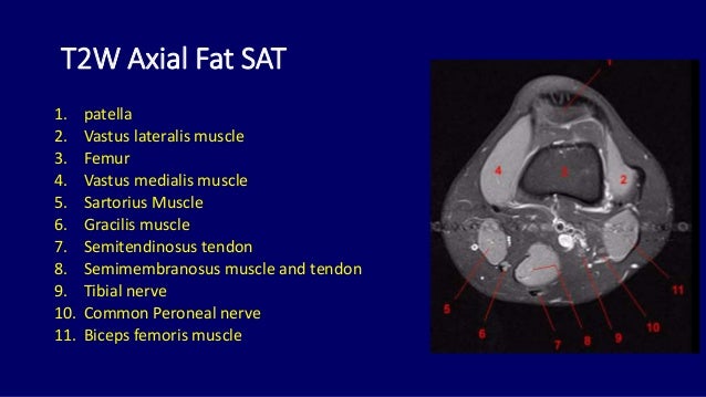
T2w axial fat sat 1.
The knee joint is a modified hinge joint between the femur tibia and patella. Injuries such as anterior cruciate ligament, meniscus and rotator cuff tears are all easily diagnosed when there is a firm understanding and knowledge of human anatomy. Intensity corresponds to a pathologic lesion. The muscles of the knee include the quadriceps, hamstrings, and the muscles of the calf. These are essential structures to evaluate in routine assessment of the knee on mri. T2w axial fat sat 1. This mri knee sagittal cross sectional anatomy tool is absolutely free to use. Plantaris can have variable size, but in most cases is difficult to demonstrate on routine mri studies. The muscles of the knee include the quadriceps, hamstrings, and the muscles of the calf. Magnetic resonance imaging mri is the imaging modality of choice in the diagnosis of acute and chronic soft tissue chondral and occult skeletal injuries of the knee. To realign the anterior cruciate ligament parallel with the sagittal imaging plane. This article is based on a presentation given by david rubin and adapted for the radiology assistant by robin smithuis. Medical images from an mri allow medical professionals to distinguish body tissues, including the meniscus (shock absorbers in the knee), cartilage, tendons, and ligaments.
This article is based on a presentation given by david rubin and adapted for the radiology assistant by robin smithuis. Magnetic resonance imaging is particularly well suited for the medical evaluation of the musculoskeletal (msk) system including the knee, shoulder, ankle, wrist and elbow. Superiorly, it extends to the level of the crossing of the biceps femoris tendon, and remains superficial to fcl in this location.10 Use the mouse scroll wheel to move the images up and down alternatively use the tiny arrows (>>) on both side of the image to move the images. In one investigation, depicted only on the proton density weighted images.
Knee Muscle Anatomy Mri : Mri Knee Anatomy Knee Sagittal ... from anatomia.wum.edu.pl Please email baodo at stanford.edu When interpreting the proton density images it. Doctors may recommend a knee mri if a patient experiences the following(3): In conclusion, we describe the normal mri anatomy of the distal biceps femoris and the relationship of this muscle with the common peroneal nerve. Use the mouse scroll wheel to move the images up and down alternatively use the tiny arrows (>>) on both side of the image to move the images. Cross sectional anatomy of the knee based on mri : Anatomy of the knee can be complicated and hard to understand. Prescribe sagittal plane off axial images with line parallel to bony glenoid.
These motions of the knee allow the body to perform such important movements as walking, running, kicking, and jumping.
In one investigation, depicted only on the proton density weighted images. The thigh has some of the body's largest muscles. These muscles work in groups to flex, extend and stabilize the knee joint. Please email baodo at stanford.edu Articular muscle of the knee (articularis genu m.) normal mr imaging anatomy of the knee. Use the mouse scroll wheel to move the images up and down alternatively use the tiny arrows (>>) on both side of the image to move the images. The images may also help physicians to distinguish normal, healthy tissues from dead tissues(2). Mri knee anatomy scroll using the mouse wheel or the arrows. Anatomy of the knee can be complicated and hard to understand. Doctors may recommend a knee mri if a patient experiences the following(3): When interpreting the proton density images it. Anatomical structures of the lower limb (hip, thigh, knee, leg, ankle and foot) and specific regions (compartment of the lower. While a detailed explanation of mri protocols and mr physics is beyond the scope of this text, fast spin echo (fse) mri is most commonly utilized for mri of the knee.
The thigh has some of the body's largest muscles. The muscles of the knee include the quadriceps, hamstrings, and the muscles of the calf. Magnetic resonance imaging mri is the imaging modality of choice in the diagnosis of acute and chronic soft tissue chondral and occult skeletal injuries of the knee. Anatomy arthrogram anatomy basic shoulder mri. Thigh muscles are responsible for allowing normal gait and proper lower extremity function (1).
Knee Mri Anatomy - Anatomy Drawing Diagram from image.slidesharecdn.com Intensity corresponds to a pathologic lesion. Medical images from an mri allow medical professionals to distinguish body tissues, including the meniscus (shock absorbers in the knee), cartilage, tendons, and ligaments. Knee muscle anatomy axial mri : Plantaris can have variable size, but in most cases is difficult to demonstrate on routine mri studies. Anatomical structures of the lower limb (hip, thigh, knee, leg, ankle and foot) and specific regions (compartment of the lower. Stanford bone tumor ddx | iss/ssr msk lectures | search ocad cases | stanford virtual readouts stanford msk mri atlas has served over 1,000,000 pages to users in over 100 countries. Use the mouse scroll wheel to move the images up and down alternatively use the tiny arrows (>>) on both side of the image to move the images.>>) on both side of the image to move the images. Magnetic resonance imaging is particularly well suited for the medical evaluation of the musculoskeletal (msk) system including the knee, shoulder, ankle, wrist and elbow.
Anatomy arthrogram anatomy basic shoulder mri.
Plantaris can have variable size, but in most cases is difficult to demonstrate on routine mri studies. Use the mouse scroll wheel to move the images up and down alternatively use the tiny arrows (>>) on both side of the image to move the images. Use the mouse scroll wheel to move the images up and down alternatively use the tiny arrows (>>) on both side of the image to move the images.>>) on both side of the image to move the images. Muscle anatomy get body smart 12 photos of the muscle anatomy get body smart muscle anatomy get body smart, human muscles, muscle anatomy get body smart The medial thigh muscles are responsible for the adduction (movement of a body part toward the body's midline) of the leg. The muscles of the knee include the quadriceps, hamstrings, and the muscles of the calf. Naturally, in order to assess pathologic knee imaging, it is necessary to know the appearance of a normal knee mri. The knee joint is a modified hinge joint between the femur tibia and patella. View of the anatomical labels. Prescribe sagittal plane off axial images with line parallel to bony glenoid. In conclusion, we describe the normal mri anatomy of the distal biceps femoris and the relationship of this muscle with the common peroneal nerve. Articular surface of patella and femur, condyle, epicondyle and muscles (popliteus, sartorius, gastrocnemius, semimembranous with tendos.) the images obtained were exported to jpeg from dicom data stored on the pacs (picture archiving and communicating system). This article is based on a presentation given by david rubin and adapted for the radiology assistant by robin smithuis.